Exploring the Human Eye: Anatomy, Function & Health
1. Eye Anatomy Overview
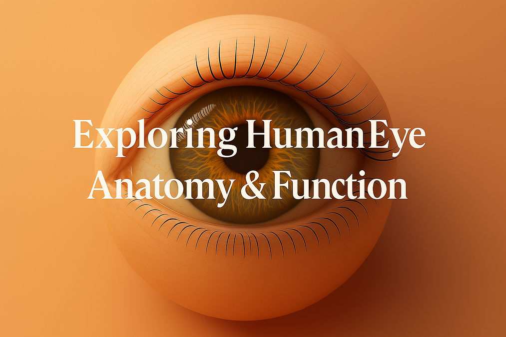
1.1 Cornea
The cornea is the clear, dome-shaped window covering the front of the eye, responsible for two-thirds of its focusing power. Composed of five layers, it refracts incoming light and protects internal structures :contentReference[oaicite:4]{index=4}.
1.2 Lens
Directly behind the iris sits the crystalline lens, a flexible, biconvex structure that fine-tunes focus via accommodation (shape changes driven by ciliary muscles) :contentReference[oaicite:5]{index=5}.
1.3 Retina
Lining the back of the eye, the retina’s light-sensitive rods and cones transduce photons into electrical impulses. Cones in the fovea support high-acuity, color vision, while rods enable low-light sight :contentReference[oaicite:6]{index=6}.
1.4 Optic Nerve
The optic nerve transmits visual signals from retinal ganglion cells to the brain’s visual cortex. It forms the natural blind spot where no photoreceptors are present :contentReference[oaicite:7]{index=7}.
2. How Vision Works
2.1 Refraction
Light entering through the cornea and lens bends (refracts) to focus on the retina. Precise corneal curvature and lens flexibility ensure sharp images. Errors in this system cause refractive issues like myopia or astigmatism :contentReference[oaicite:8]{index=8}.
2.2 Neural Processing
After phototransduction, signals pass through bipolar and ganglion cells, cross at the optic chiasm, and reach the visual cortex. The brain integrates input from both eyes for depth perception and image clarity :contentReference[oaicite:9]{index=9}.
3. Frequently Asked Questions
Iris melanin concentration dictates hues from blue to brown.
The iris adjusts pupil size to regulate light entry and protect the retina.
Adults should test vision every 1–2 years; children by age 1, 3, and before school.
Protein clumping in the lens, often age-related, leads to clouded vision.
Retinal cells have limited regeneration; damage can be permanent.
Increased intraocular pressure damages the optic nerve, potentially causing blindness.
Vitreous gel clumps casting shadows on the retina, usually harmless but should be monitored.
Laser reshapes the cornea to correct refractive errors, reducing dependence on glasses.
Prolonged use leads to digital eye strain; follow the 20-20-20 rule (every 20 min, look 20 ft away for 20 sec).
Vitamins A, C, E, zinc, lutein, and omega-3 fats promote retinal function.
Increased outdoor activity and controlled near work can slow progression in children.
Often X-linked, it results from cone photopigment anomalies affecting color discrimination.
Tears lubricate, nourish, and protect the ocular surface from irritants.
Age-related lens stiffening reduces near-focus ability, requiring reading glasses.
Wear UV-blocking sunglasses and broad-brim hats outdoors.
Join the conversation